A polyp can grow for years without causing bleeding, pain or a change in bowel habits, yet some types can eventually become colorectal cancer. By understanding which lesions matter, who needs screening, and what happens after an abnormal result, you can act before a precancerous growth becomes a cancer that requires more extensive treatment.
Key takeaways
- Remove precancerous polyps before they invade the bowel wall.
- Treat size, number and microscopic features as key risk clues.
- Use FIT for stool screening; colonoscopy finds and removes polyps.
- Keep every recommended follow-up after an abnormal result.
How a normal colon lining can become a cancerous growth
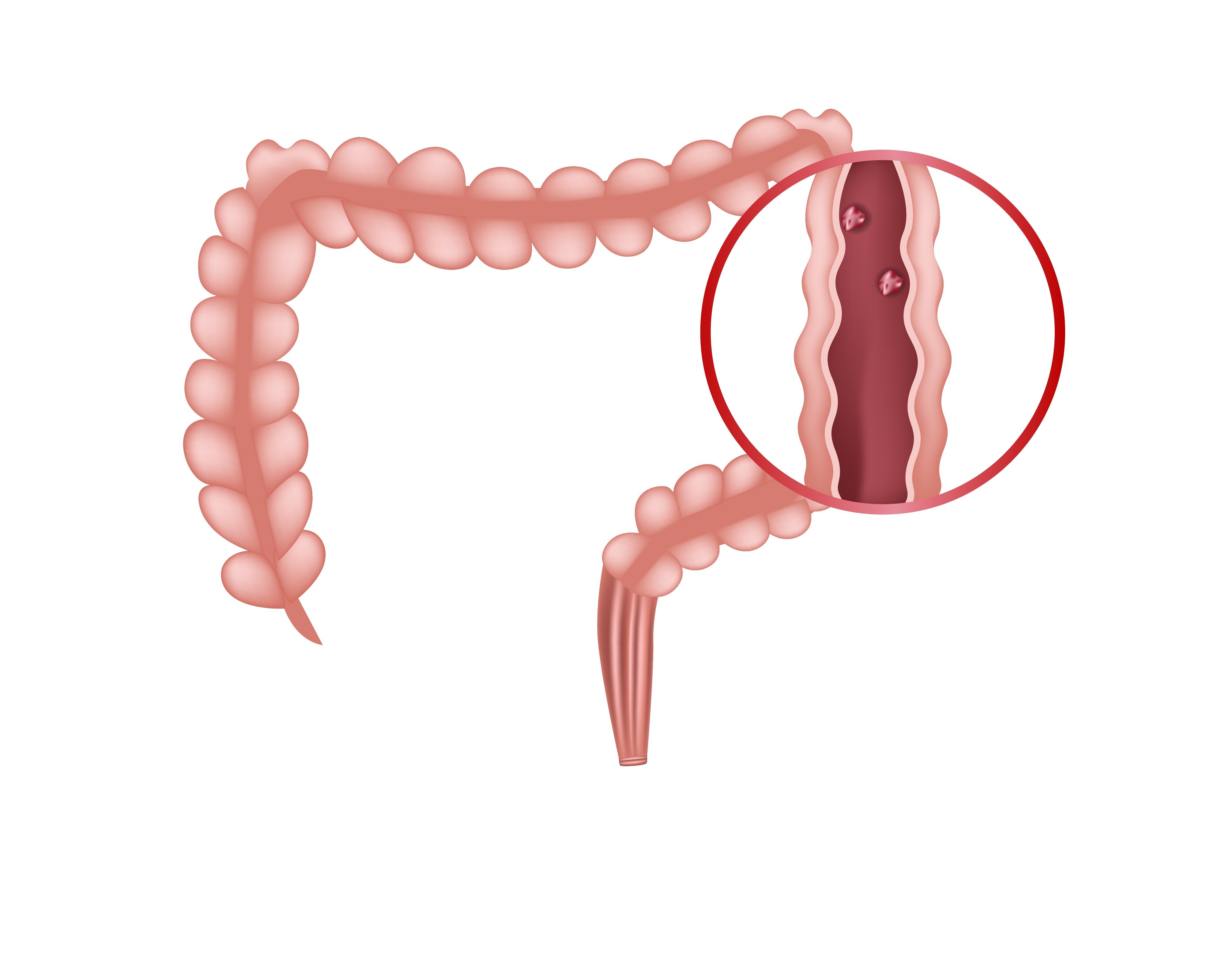
A colorectal polyp begins when cells in the normal colon lining grow and divide abnormally, creating a bump or flat area. Most remain harmless, but some collect genetic changes that let them grow larger, invade nearby tissue and eventually become colorectal cancer.
The best-known route is the adenoma–carcinoma sequence. An adenomatous polyp develops first, then accumulates cellular changes over roughly 10–15 years before becoming cancerous; that timetable is not guaranteed, and removal can interrupt it. A separate serrated pathway begins with serrated polyps, which can be flat, subtle and located on the right side of the colon.
| Type | Cancer potential | What to know |
|---|---|---|
| Adenomatous polyps | Precancerous | Tubular, villous and tubulovillous adenomas can progress, with risk influenced by size and microscopic features. |
| Serrated polyps | Some are precancerous | Sessile serrated lesions and traditional serrated adenomas can lead to cancer through the serrated pathway and are easier to miss when flat. |
| Hyperplastic polyps | Usually little or no risk | Small hyperplastic polyps in the rectosigmoid colon are generally not precancerous. |
| Inflammatory or other growths | Usually not direct precursors | Their meaning depends on the diagnosis; inflammatory bowel disease itself raises colorectal cancer risk. |
A polyp’s number, size, location and pathology determine how seriously it should be followed. Finding and completely removing a precancerous lesion prevents that lesion from reaching cancer; detecting an established cancer is a different goal.
Why a polyp’s size, number and microscopic features change the risk
An 8-mm tubular adenoma removed completely is a different risk finding from a 15-mm adenoma removed in pieces. Polyp size reflects the time and opportunity for abnormal cells to accumulate, while pathology shows whether the cells have villous features or high-grade dysplasia—changes linked with greater progression risk.
The number of polyps also matters. Multiple adenomas suggest a colon that is producing precancerous growths more actively, so surveillance needs to be sooner. Location adds context: a small hyperplastic polyp in the rectosigmoid colon usually has little malignant potential, while certain serrated lesions in the proximal colon can carry meaningful risk.
Under U.S. Multi-Society Task Force guidance, common surveillance patterns include:
- One or two tubular adenomas smaller than 10 mm: repeat colonoscopy in 7–10 years.
- An adenoma at least 10 mm, with villous features, or with high-grade dysplasia: repeat in 3 years.
- Five to 10 adenomas: repeat in 3 years; more than 10: repeat in 1 year.
- A lesion at least 20 mm removed piecemeal: recheck in about 6 months.
Complete removal is as important as the laboratory diagnosis. Tissue left behind can grow again, and piecemeal removal makes it harder to confirm a clear edge. The endoscopy report and pathology report must therefore be read together before setting the next examination.
Screening before symptoms versus testing because symptoms are present
Average-risk adults should discuss colorectal cancer screening from age 45 through 75. Screening without symptoms looks for precancerous polyps before bleeding, bowel-habit changes, pain or weight loss appear. From 76 through 85, decide selectively with a clinician by weighing overall health, previous screening and life expectancy.
Discuss an earlier or more intensive plan if you have:
- A first-degree relative with colorectal cancer or advanced polyps
- Inflammatory bowel disease involving the colon
- Lynch syndrome, familial adenomatous polyposis or another hereditary cancer syndrome
- A previous adenoma or colorectal cancer
Screening and diagnostic colonoscopy answer different questions.
| Purpose | Starting point | What the colonoscopy means |
|---|---|---|
| Screening | No symptoms; a scheduled test such as FIT or screening colonoscopy | Looks for cancer or precancerous lesions before they cause trouble |
| Diagnostic colonoscopy | Symptoms such as rectal bleeding, or an abnormal FIT or other test | Investigates a warning sign and may biopsy or remove a lesion |
A positive stool-based screening test is not proof of cancer or even a polyp. It requires follow-up diagnostic colonoscopy; skipping that step removes much of the test’s potential benefit.
Colonoscopy can cause bleeding, perforation, sedation reactions or bowel-preparation complications, with risk rising after polyp removal and with age or frailty. Your clinician should balance those harms against the benefit of removing a precancerous polyp before it becomes cancer.
FIT or colonoscopy: what early detection requires from each test
A faecal immunochemical test (FIT) asks you to collect a small stool sample at home and send it for hidden human blood. It usually needs no fasting, sedation or colonoscopy preparation, but it detects bleeding rather than polyps themselves. Bleeding can be intermittent, and a negative result does not exclude a non-bleeding lesion.
| Option | Preparation | Follow-up and limitation |
|---|---|---|
| FIT | Collect stool at home as instructed; no bowel cleansing | Repeat at the screening interval. A positive stool test requires colonoscopy; FIT cannot remove a polyp or explain the bleeding. |
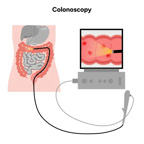
| Colonoscopy | Follow the prescribed low-residue diet, clear-liquid period and split-dose laxative; arrange transport if sedation is used | The endoscopist examines the whole colon and can biopsy or remove precancerous polyps during the same procedure, preventing that lesion from progressing to cancer. |
A positive FIT is not a cancer diagnosis or proof of a polyp. Delaying the follow-up colonoscopy removes much of FIT’s preventive value.
Colonoscopy offers the more complete single examination, but it is not risk-free: bowel-preparation complications, sedation reactions, bleeding and perforation are possible. Risk rises after polyp removal and in older or medically frail people.
A normal result is not an absolute guarantee; poor preparation, flat or right-sided lesions and rushed withdrawal inspection can lead to missed growths. Ask how preparation quality and complete examination were documented, then follow the recommended surveillance interval.
Turning an abnormal finding into lasting cancer prevention
A positive stool test is not a cancer diagnosis, but it does require a follow-up diagnostic colonoscopy. Without that examination, a potentially preventable cancer or polyp can remain undiscovered.
After a polyp is found, keep the pathology report and procedure report together. Ask whether removal was complete, when the next surveillance colonoscopy is due, and which finding sets the interval.
- Five to 10 adenomas generally call for colonoscopy in three years.
- More than 10 adenomas generally call for colonoscopy in one year.
- Piecemeal removal of a lesion at least 20 mm calls for a site-check colonoscopy in about six months.
- Incomplete polyp removal needs a documented plan for completing treatment or checking the remaining tissue.
Piecemeal removal prevents the endoscopist from confirming complete excision from the specimen alone. Do not treat a later appointment as optional.
Personal risk changes the pathway. Tell the specialist about a first-degree relative with colorectal cancer or advanced polyps, inflammatory bowel disease, previous adenomas or colorectal cancer, and any family pattern of early or multiple cancers. Lynch syndrome and familial adenomatous polyposis call for genetic evaluation and specialist-led screening plans.
Dr Pravish Ghurde Gastroenterologist can help interpret pathology alongside removal quality, family history and bowel inflammation when a standard interval does not fit. Balance each procedure’s benefit against bleeding, perforation, sedation and bowel-preparation risks, particularly with increasing age or frailty.
Related service
Colorectal Polyps, Lower GI Bleeding & Liver Disease Treatment Colorectal Polyps, Lower GI Bleeding & Liver Disease Treatment Dr. Pravish Ghurde, Consultant Gastroenterologist at Platinum Hospital, Mulund, Mumbai, provides expert diagnosis and advanced treatment for... View service → |
Frequently asked questions
How can a normal colon lining become a cancerous growth?
Cells can grow abnormally, form a polyp and collect genetic changes that allow invasion into nearby tissue.
Why do a polyp’s size, number and microscopic features matter?
Larger or multiple polyps and high-risk microscopic features indicate a greater chance of future colorectal cancer.
Should you screen before symptoms appear?
Yes. Screening can find precancerous polyps before symptoms develop, while diagnostic testing investigates symptoms that are already present.
What is the difference between FIT and colonoscopy for early detection?
FIT checks stool for hidden blood, while colonoscopy directly examines the colon and allows the doctor to remove or biopsy polyps.
What should you do after an abnormal polyp or screening result?
Complete the recommended colonoscopy, pathology review and follow-up interval so detected risk leads to lasting prevention.