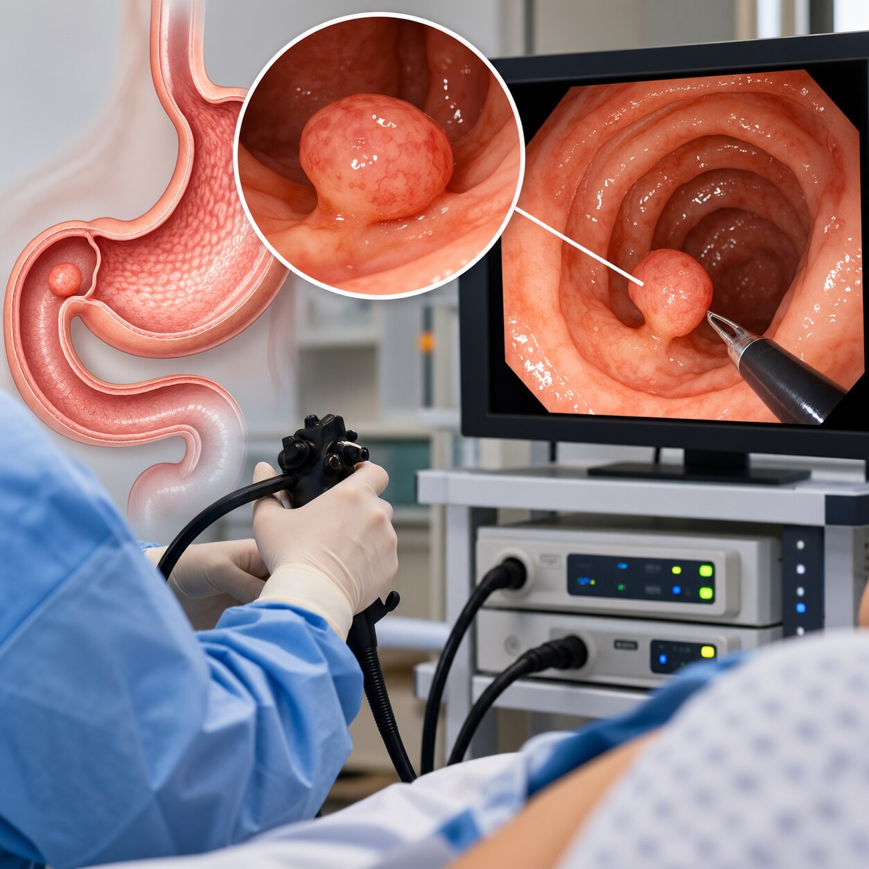
A duodenal polyp is an endoscopic finding, not a final diagnosis: the same raised area may represent an adenoma, Brunner gland hamartoma, neuroendocrine tumour, ectopic tissue, or another neoplasm. By the end, you will know how doctors locate and classify these lesions, when biopsy or removal is appropriate, and why ampullary lesions and multiple adenomas require a different plan.
Key takeaways
- A duodenal polyp describes tissue growth, not its final diagnosis.
- Doctors map the major papilla before sampling or removing nearby lesions.
- Ampullary polyps require separate assessment because bile and pancreatic ducts open there.
- Follow-up depends on pathology, complete removal, recurrence, and polyposis risk.
What a Duodenal Polyp Can Represent
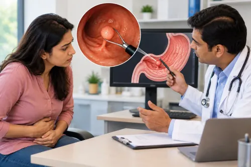
A duodenal polyp is an endoscopic description, not a diagnosis: it is tissue projecting into the duodenal lumen. A duodenal bulb polyp lies in the first portion, while a second part of duodenum polyp lies beyond it, near the major papilla.
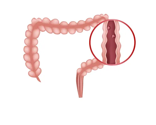
A gastric polyp arises in the stomach; an ampullary polyp involves the papilla where the bile and pancreatic ducts open; a colorectal polyp arises in the colon or rectum. Colour and shape alone cannot establish tissue type, depth, dysplasia, or duct involvement.
| Possible diagnosis | What it represents |
|---|---|
| Duodenal adenoma | A potentially precancerous mucosal lesion |
| Brunner gland hyperplasia or hamartoma | A benign glandular proliferation |
| Inflammatory polyp | Reactive tissue linked to irritation or inflammation |
| Neuroendocrine tumour | A tumour arising from neuroendocrine cells |
| Lipoma | A fatty subepithelial lesion |
| Ectopic pancreas | Pancreatic tissue in an abnormal location |
| Other subepithelial or neoplastic lesions | Conditions requiring depth assessment and pathology |
Concern rises with:
- Increasing size
- Multiple polyps
- An irregular or nodular surface pattern
- Ulceration
- Spontaneous bleeding
- A firm texture
- Proximity to the major papilla, where duct involvement can change management
How Endoscopy Locates the Lesion and the Major Papilla
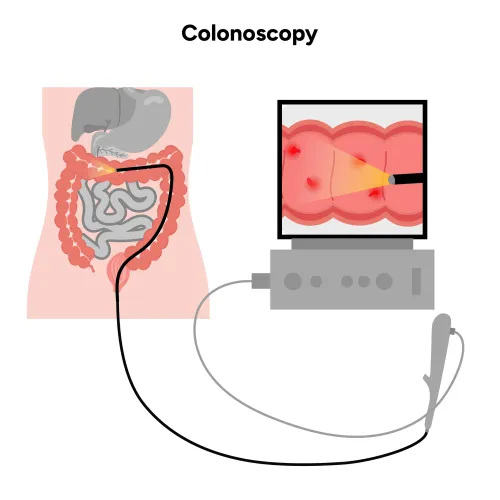
A forward-viewing gastroscope inspects the stomach, passes through the duodenal bulb, and follows the second part of the duodenum for visible polyps, ulcers, bleeding, and narrowing. Its straight-ahead view does not reliably show the major papilla because the papilla faces laterally and folds can obscure its opening.
Use a side-viewing duodenoscope when a lesion lies near the papilla. It shows whether the growth is ampullary, periampullary, or separate from the papilla and permits a search for additional periampullary disease.
- For a mucosal-appearing lesion, assess its surface, base, size, and distance from the major papilla.
- For a subepithelial-appearing lesion, use endoscopic ultrasound before snaring or repeated superficial forceps sampling.
- Confirm whether it arises from the mucosa, submucosa, or muscularis propria.
- Treat deep origin, irregular borders, ulceration, duct extension, or other invasive features as warning signs against routine snare removal.
Bile-duct or pancreatic-duct involvement changes the assessment because the lesion may extend beneath the visible surface, obstruct drainage, or require duct mapping and a different treatment plan. Endoscopic ultrasound and magnetic-resonance cholangiopancreatography can define that relationship before removal.
When Doctors Biopsy, Remove, or Wait for More Information
A clearly resectable suspected nonampullary adenoma usually belongs in removal rather than routine sampling. ESGE advises against a duodenal polyp biopsy when its result would not change the decision to resect; biopsy-induced fibrosis can make later endoscopic removal harder.
| Option | Decision | Main reason and limitation |
|---|---|---|
| Standard forceps biopsy | Sample first | A forceps biopsy can miss the lesion’s most advanced area, underestimate duodenal dysplasia, or fail to detect invasion. |
| Immediate endoscopic removal | Remove now | Appropriate for a small, clearly mucosal and resectable lesion when complete removal is feasible and biopsy would not alter management. |
| Deferred treatment | Investigate first | Appropriate when depth, tissue type, ampullary involvement, or invasive cancer remains uncertain. |
Do not automatically snare a lesion that looks subepithelial, has uncertain depth, lies near the major papilla, or shows ulceration, firmness, spontaneous bleeding, or other invasive features. Endoscopic ultrasound can show whether it arises from the mucosa, submucosa, or muscularis propria; cross-sectional imaging and duct evaluation can define spread.
Pathology must answer three questions: is this an adenoma or another lesion, such as a neuroendocrine tumour or Brunner-gland lesion; is the dysplasia low-grade or high-grade; and is there evidence of invasion? Those findings determine whether removal, staging, surveillance, or surgery comes next.
Choosing a Resection Method and Preventing Complications
Size, morphology, lifting, depth, and location determine duodenal polyp removal. A small, smooth, superficial lesion away from the papilla may suit cold-snare removal; a larger or thicker stalked lesion may need conventional snare polypectomy. Failure to lift, deep involvement, or proximity to the major papilla changes the plan.
| Option | Best fit | Main limitation |
|---|---|---|
| Cold-snare removal | Small, superficial, nonampullary lesions | Limited use for thick or broad lesions |
| Conventional snare polypectomy | Pedunculated or visibly resectable lesions | Thermal injury and bleeding risk |
| Endoscopic mucosal resection | Larger nonampullary adenomas without clear invasive cancer | Broad lesions often require piecemeal removal |
| Endoscopic submucosal dissection | Selected lesions needing en-bloc, precise excision | High perforation and bleeding risk in the duodenum |
Endoscopic mucosal resection is usually first-line for a larger nonampullary adenoma without clear invasive cancer. Piecemeal removal of a broad lesion limits lateral and deep margin assessment and increases local-recurrence risk.
Endoscopic submucosal dissection is reserved for selected cases in highly experienced centres because the thin duodenal wall and rich blood supply raise perforation and bleeding risks.
The endoscopist often closes the post-resection defect with clips to reduce delayed bleeding and perforation risk. Seek urgent care for severe pain, vomiting blood, black stools, fever, or faintness after the procedure.
Why Ampullary Polyps Need a Separate Treatment Pathway
An ampullary polyp cannot be treated like an ordinary nonampullary polyp because it arises where the bile and pancreatic ducts empty. An ampullary adenoma can obstruct drainage or extend inside either duct, while routine snaring cannot map this anatomy or exclude invasive cancer.
A side-viewing duodenoscope defines the major papilla and the lesion’s relationship to it. Targeted biopsies sample suspicious tissue, while endoscopic ultrasound and/or magnetic-resonance cholangiopancreatography provide bile duct and pancreatic duct evaluation, assess depth, and detect intraductal extension or invasion.
Endoscopic papillectomy is considered when staging shows no convincing invasive cancer or significant duct extension. Surgery may be preferred when invasion or substantial duct involvement is present because endoscopic removal could leave cancer behind or fail to clear the ducts.
Before papillectomy, discuss these safeguards and complications:
- Place a prophylactic pancreatic-duct stent when appropriate, especially after pancreatic-duct instrumentation or opacification.
- Give rectal nonsteroidal anti-inflammatory medication unless contraindicated; neither measure eliminates pancreatitis.
- Monitor for immediate or delayed bleeding, perforation, pancreatitis, cholangitis, and later papillary stenosis.
Follow-Up After Removal and Assessment for Polyposis Syndromes
An apparently complete removal does not end duodenal polyp surveillance. ESGE recommends inspection about 3 months after duodenal adenoma removal, followed by another examination at 1 year when no residual or recurrent lesion is found. Later intervals depend on recurrence, histology, and completeness of removal.
| Finding | Broader evaluation | Management implication |
|---|---|---|
| Single adenoma | Review pathology and resection margins | Individualise duodenal adenoma follow-up |
| Multiple adenomas | Examine the colon and remainder of the gastrointestinal tract; review three generations of family history | Assess for familial adenomatous polyposis or another polyposis syndrome |
| Familial adenomatous polyposis | Stage upper-GI disease with the Spigelman classification | Adjust surveillance and treatment intensity |
The Spigelman classification uses the number, size, histology, and dysplasia of duodenal adenomas. A higher stage means closer surveillance and consideration of more intensive treatment.
For complex biopsy, resection, or bile- and pancreatic-duct assessment, Dr Pravish Ghurde Gastroenterologist can coordinate diagnosis, pathology review, surveillance, and referral for surgery when endoscopic treatment is unsuitable.
Frequently asked questions
What can a duodenal polyp represent?
A duodenal polyp is tissue projecting into the duodenal lumen. Its appearance can represent different growths, so pathology may be needed for diagnosis.
How does endoscopy locate a duodenal polyp and the major papilla?
Upper endoscopy examines the duodenal bulb and second portion while documenting the lesion’s size, surface, location, and relationship to the major papilla.
When is a duodenal polyp biopsied, removed, or observed?
Doctors choose among biopsy, resection, or short-term observation based on the lesion’s appearance, size, location, bleeding risk, and the information needed from pathology.
Why do ampullary polyps need a separate treatment pathway?
The major papilla is where the bile duct and pancreatic duct enter the duodenum. Treatment must account for duct involvement and the risk of pancreatitis, bleeding, or impaired drainage.
What follow-up is needed after duodenal polyp removal?
Follow-up is guided by the pathology report, whether removal was complete, recurrence risk, and signs of a polyposis syndrome. Some patients need repeat endoscopy or genetic assessment.