A bleeding varix needs more than an endoscopic band: clinicians must stabilize circulation, reduce portal pressure, identify the varix pattern, and prevent another haemorrhage. By the end, you will know when variceal band ligation is appropriate, what the procedure involves, how it compares with other treatments, and what follow-up protects against rebleeding.
Key takeaways
- EVL places tight rubber bands around bleeding-prone oesophageal varices.
- Active bleeding needs urgent endoscopic treatment and hospital monitoring.
- Gastric varices often require different treatments because of their location.
- Follow-up endoscopy and portal-pressure treatment help prevent rebleeding.
Why oesophageal and gastric varices bleed
Cirrhosis scars the liver and obstructs blood flowing through it, raising pressure in the portal vein. This portal hypertension redirects blood through smaller veins in the oesophagus and stomach, causing oesophageal varices and gastric varices to enlarge.
These veins have thin walls and carry blood under increased pressure. A tear can therefore release blood rapidly, causing cirrhosis-related bleeding with vomiting blood or black, tarry stools. Treat those symptoms as an emergency requiring immediate assessment.
Oesophageal varices usually run along the lower oesophagus and are commonly reached for endoscopic banding. Gastric varices lie in the stomach, where their size, depth and connections to other veins can require a different treatment.
| Location | Typical implication | Why endoscopy matters |
|---|---|---|
| Oesophageal varices | Often suitable for band ligation | Confirms the bleeding point and varix size |
| Gastric varices | May need injection, coils or radiological treatment | Defines the pattern and connected veins |
| Non-variceal source | Ulcer, tear or another lesion | Requires treatment directed at that cause |
Upper endoscopy is essential because vomiting blood or black stools do not prove that a varix is responsible. Variceal band ligation is intended for a confirmed oesophageal-variceal source or high-risk varices, not every case of upper gastrointestinal bleeding. It controls the bleeding point but does not cure portal hypertension or cirrhosis.
How variceal band ligation stops oesophageal bleeding
Variceal band ligation, also called endoscopic variceal ligation or EVL, treats a confirmed oesophageal varix rather than every cause of oesophageal bleeding. An endoscope identifies the bleeding point or a varix with high-risk features before treatment begins.
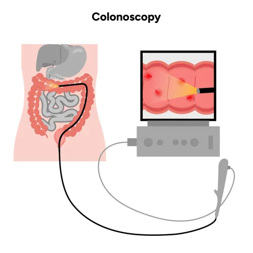
- The clinician passes the endoscope through the mouth into the oesophagus and positions its tip over the varix.
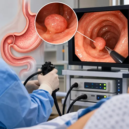
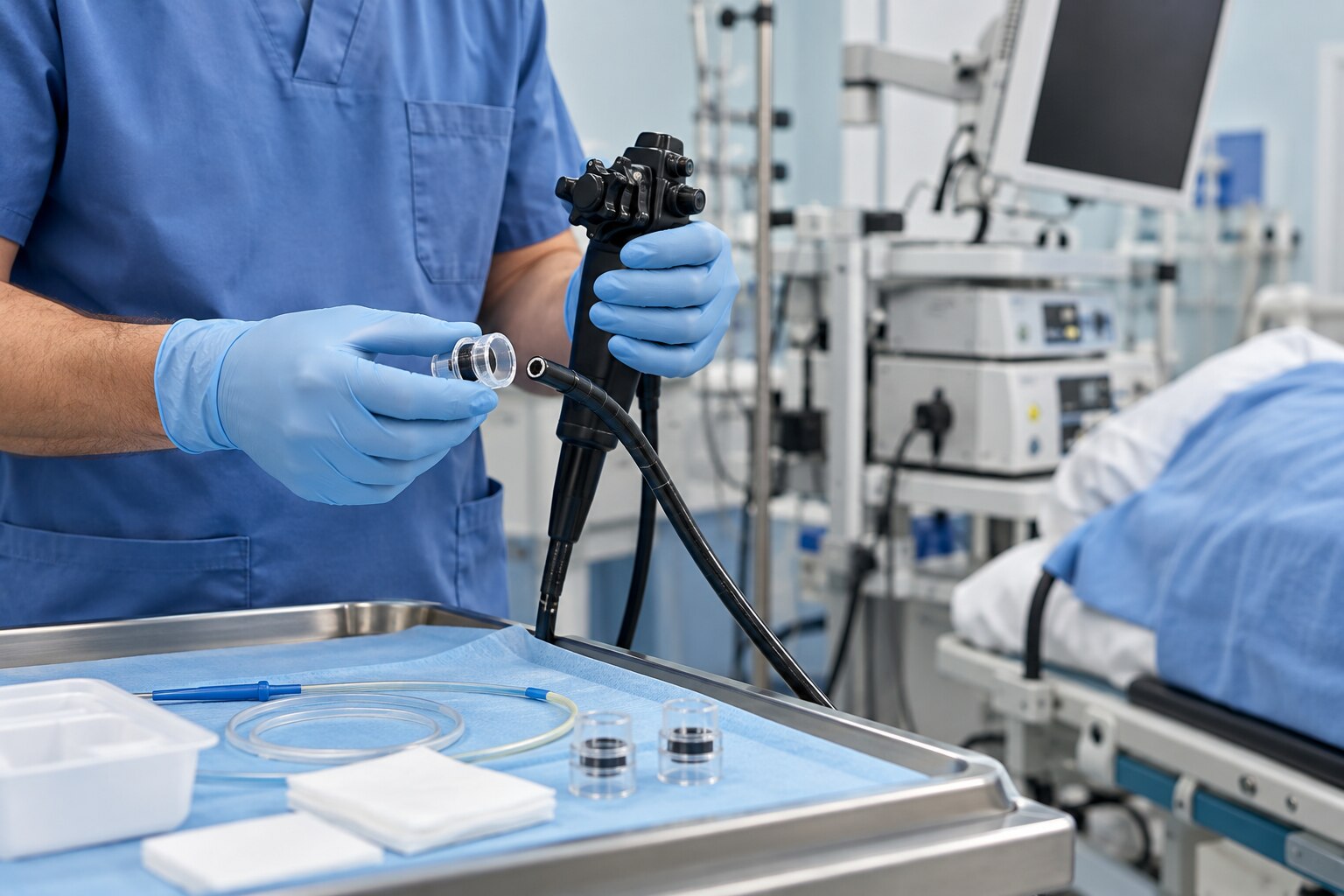
- Suction draws the bleeding or high-risk varix into a transparent cap attached to the endoscope.
- A small elastic band is released around the varix’s base, tightly closing the vessel and cutting off its blood flow.
- The trapped tissue shrinks and detaches, leaving an ulcer underneath that heals as the area recovers.
Active haemorrhage requires immediate control. High-risk varices found before bleeding are treated to prevent a first bleed in selected patients. Planned repeat EVL sessions, commonly every 1–4 weeks, remove residual varices until eradication; surveillance follows because varices can return.
For primary prevention, EVL is an option for selected medium or large varices when a nonselective beta blocker is contraindicated, unsuitable or not tolerated. Every small varix does not require banding.
Chest discomfort, painful swallowing and temporary dysphagia are common short-term effects. Vomiting blood, black stools, faintness or marked weakness needs emergency care because a post-banding ulcer can bleed days later.
What happens when a varix is actively bleeding
An actively bleeding varix requires an emergency bundle, not banding alone. Establish intravenous access, assess circulation and airway, and test haemoglobin, platelet count and clotting studies while preparing urgent endoscopy.
- Restore circulation without overloading the patient.
- Consider red-cell transfusion around a haemoglobin of 7 g/dL, usually targeting 7–9 g/dL; liberal transfusion can raise portal pressure and increase rebleeding risk.
- Start a vasoactive drug such as octreotide or terlipressin to reduce splanchnic blood flow.
- Give antibiotic prophylaxis to patients with cirrhosis, even without obvious infection. Treatment often continues for up to seven days, guided by local resistance patterns and individual risk.
- Arrange endoscopy after initial stabilisation, generally within 12 hours, so the bleeding source can be confirmed and oesophageal variceal banding performed.
A high-risk patient can need early or pre-emptive transjugular intrahepatic portosystemic shunt (TIPS), ideally within 24 hours and no later than 72 hours after endoscopy. This includes selected Child-Pugh class C patients with a score below 14 and selected Child-Pugh class B patients with active bleeding despite vasoactive treatment.
TIPS does not replace urgent endoscopy and banding; it reduces portal pressure when the risk of treatment failure or rebleeding remains high.
Why the varix location changes the treatment choice
Varix location changes treatment because anatomy determines whether a band can grasp the vessel and whether portal pressure can be redirected. For oesophageal varices, EVL is generally preferred to injection sclerotherapy: mechanical control achieves haemostasis with fewer treatment-related complications. Sclerotherapy remains useful when band placement is technically impossible.
| Option | Best fit | Main limitation |
|---|---|---|
| EVL | Oesophageal varices and selected gastroesophageal patterns | Cannot reliably treat many gastric varices |
| Cyanoacrylate or thrombin injection | Isolated gastric varices and suitable gastric variceal bleeding | Requires careful delivery and expertise |
| EUS-guided coil or glue therapy | Gastric varices with accessible feeding vessels | Availability and anatomy limit its use |
| Radiologic shunt or obliteration | Gastric patterns unsuitable for endoscopic treatment | Requires specialised imaging and procedural teams |
Balloon tamponade or a self-expanding oesophageal stent provides temporary rescue control when endoscopic treatment fails; neither is a definitive cure. TIPS decompresses the portal system for uncontrolled or recurrent bleeding and may be considered in selected high-risk cases.
A band closes the bleeding point, not the disease causing it. It does not cure portal hypertension or cirrhosis, so varices can recur and require surveillance and treatment of the underlying liver disease.
Preparing for EVL and knowing what recovery involves
An active bleed usually requires hospital admission for stabilization and urgent endoscopy. Preventive EVL can be a planned outpatient procedure when you are stable, but local unit rules and your condition determine admission, transport, procedure length, and recovery time.
- Follow the fasting before endoscopy instructions exactly, including when to stop food, milk, and clear fluids.
- Bring a complete medication list and discuss anticoagulants, antiplatelet medicines, diabetes treatment, allergies, and liver disease. Do not stop prescribed medicines unless the treating team tells you to.
- Review the consent form, arrange intravenous access, and agree on sedation or anaesthesia. Sedation usually makes you drowsy and relaxed; the endoscope can cause pressure, gagging, or brief throat discomfort.
After banding, chest discomfort, painful swallowing, and temporary difficulty swallowing can occur. Arrange a responsible adult to accompany you home after sedation, and follow the unit’s restrictions on driving and important decisions.
Fresh blood in vomit, black stools, faintness, or severe weakness requires emergency care. A post-banding ulcer can bleed several days later.
Preventing rebleeding after the bands are placed
After an episode of variceal bleeding, secondary prevention usually combines repeat EVL sessions with a nonselective beta blocker, unless the medicine is contraindicated or not tolerated. EVL alone is inferior because it closes the bleeding point but does not lower portal pressure.
Sessions are commonly repeated every 1–4 weeks until the oesophageal varices are eradicated. Endoscopic surveillance continues afterward because new or recurrent varices can develop. Treating the cause of portal hypertension and the underlying liver disease remains essential; a band is not a permanent cure.
Ask your gastroenterologist:
- Where are the varices, and what is my Child-Pugh risk?
- Is a nonselective beta blocker safe with my blood pressure, kidney function and other medicines?
- When is my next endoscopy, and how many repeat EVL sessions might I need?
- Do I need antibiotics now, and what determines the choice and duration?
- What findings would trigger a TIPS referral?
Dr Pravish Ghurde Gastroenterologist in Mulund can help assess these factors and coordinate a plan suited to your liver disease, bleeding risk and treatment tolerance.
Related services
Upper GI Bleeding Treatment Dr. Pravish Ghurde provides prompt and advanced care for upper gastrointestinal (GI) bleeding at Platinum Hospital, Mulund, Mumbai. View service → |
Colorectal Polyps, Lower GI Bleeding & Liver Disease Treatment Colorectal Polyps, Lower GI Bleeding & Liver Disease Treatment Dr. Pravish Ghurde, Consultant Gastroenterologist at Platinum Hospital, Mulund, Mumbai, provides expert diagnosis and advanced treatment for... View service → |
Frequently asked questions
Why do oesophageal and gastric varices bleed?
Cirrhosis can cause portal hypertension, redirecting blood into enlarged veins in the oesophagus and stomach. Their thin walls can rupture under pressure.
How does variceal band ligation stop oesophageal bleeding?
An endoscope places a small rubber band around the varix. The band cuts off blood flow, allowing the treated tissue to shrink and heal.
What happens when a varix is actively bleeding?
Active bleeding requires urgent hospital care, stabilisation, and endoscopic treatment. The medical team controls bleeding while treating the underlying portal hypertension.
Why does varix location change the treatment choice?
Oesophageal varices are often accessible for band ligation. Gastric varices have different blood flow and anatomy, so another endoscopic or radiological treatment may be more suitable.
What does recovery after EVL involve?
You may have temporary chest discomfort or difficulty swallowing. Follow the discharge instructions, take prescribed medicines, and attend planned follow-up endoscopies.
Related services
Ulcerative Colitis Treatment Ulcerative Colitis Treatment in Mulund, Mumbai by Dr. Pravish Ghurde Dr. View service → |
Gastroparesis Treatment Dr. Pravish Ghurde provides specialized diagnosis and comprehensive treatment for gastroparesis at Platinum Hospital, Mulund, Mumbai. View service → |