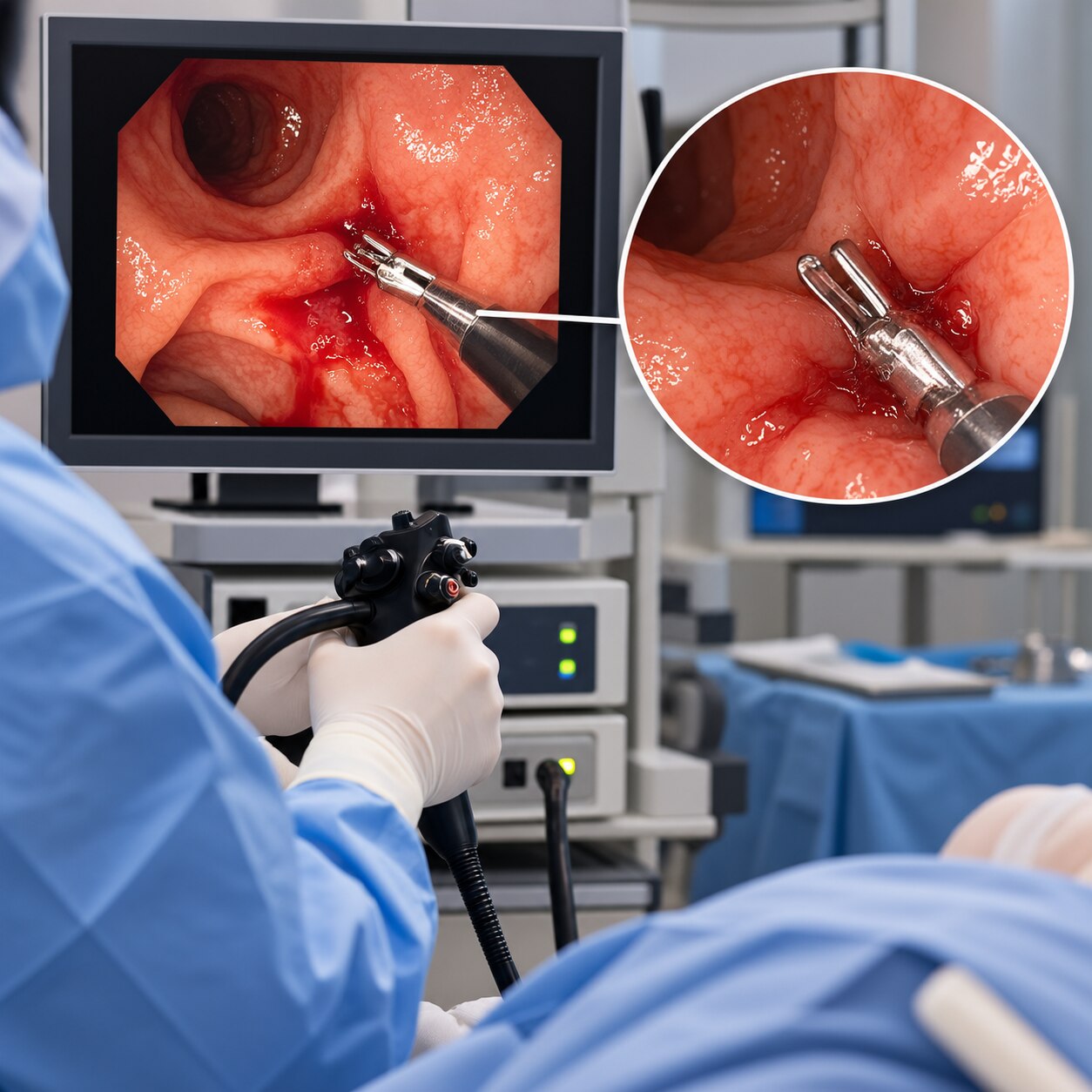
A hemoclip lets an endoscopist compress a bleeding vessel or its surrounding tissue from inside the digestive tract, without an external incision. By the end, you will understand how the clip is delivered, which bleeding findings it can treat, how technical success is confirmed, and what happens when clipping is not enough.
Key takeaways
- Hemoclips reach bleeding through the endoscope’s working channel.
- Endoscopists match clip design to the vessel, lesion, and access angle.
- Clipping may be combined with injection or thermal treatment.
- A retained clip is usually assessed alongside symptoms and rebleeding signs.
How an Endoscopist Delivers a Hemoclip to the Bleeding Point
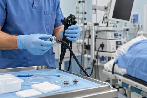
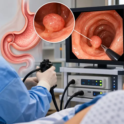
A hemoclip reaches the bleeding point through the endoscope working channel, so the endoscopist can treat the vessel without an external incision. After guiding the flexible endoscope to the stomach, colon, or another gastrointestinal segment, the endoscopist suctions blood and washes away clot to expose the source.
The target is the culprit vessel or the tissue around it, not simply the visible pool of blood.
1. The endoscopist advances a clip applicator through the endoscope working channel and positions its tip close to the bleeding point.
2. The clip opens at the tip. The endoscopist adjusts the endoscope and applicator until the jaws approach the vessel from a stable, en-face angle.
3. The jaws capture the vessel or the tissue immediately over it. Firm closure produces bleeding vessel compression, pinching the vessel shut.
4. The endoscopist confirms that bleeding has stopped before releasing the clip. More than one clip may be placed when the vessel is broad or the first clip does not fully compress it.
This is mechanical hemostasis: the clip closes tissue by pressure rather than delivering heat. That distinction helps limit thermal injury to nearby bowel or stomach tissue. If blood obscures the target or the endoscopist cannot obtain a stable approach, accurate clip placement becomes difficult and another treatment method may be needed.
Which Bleeding Findings Can Hemoclips Treat—and Which Clip Design Fits?
A hemoclip treats a focal bleeding point by compressing the culprit vessel or the tissue immediately around it. Suitable findings include a spurting ulcer, oozing ulcer, non-bleeding visible vessel, Mallory-Weiss tear, bleeding polyp stalk, post-polypectomy site, diverticular hemorrhage, and selected lesions in the colon or small bowel.
A clean ulcer base or flat pigmented spot usually needs no endoscopic hemostasis.
Clip design determines how accurately and deeply the target can be captured.
| Option | What it means | When it applies |
|---|---|---|
| Standard through-the-scope clip | Opens and closes through the endoscope, with limited control of orientation and repositioning | Accessible vessels, small tears, polyp stalks, and straightforward ulcer bleeding |
| Rotatable clip | The endoscopist can turn the clip before closing it | A vessel needs a particular clip angle, especially when the approach is not directly face-on |
| Repositionable clip | The clip can be opened, adjusted, and reopened before release | Precise placement is difficult, or the first position does not capture the vessel |
| Over-the-scope clip | A larger cap-mounted clip captures a substantial, deeper tissue bite | Large-caliber arteries, fibrotic ulcers, larger defects, or bleeding that continues after standard clips |
Standard clips can close over blood without grasping the artery when the target lies deep, behind a fold, or at a tangential angle. Diverticular necks and thin-walled tissue also demand careful placement; an over-the-scope clip is stronger, but it requires deliberate tissue capture and is not a routine substitute.
When Clipping Is Used Alone, Combined With Other Treatment, or Avoided
A hemoclip provides definitive hemostasis when it fully compresses an accessible bleeding vessel or nonbleeding visible vessel and bleeding stops after inspection. For peptic ulcers, clipping alone can be sufficient for a focal spurting point, oozing point, or visible vessel that the endoscopist can grasp securely.
A clean ulcer baseurcret, meaning a clean ulcer base, and a flat pigmented spot do not need endoscopic hemostasis. Treating them adds manipulation without a target to close. A broad ulcer surface, diffuse oozing, or a vessel hidden by an adherent clot can defeat a focal clip.
Dilute epinephrine is an aid, not definitive hemostasis. Injecting it around a bleeding ulcer slows flow and clears the view; the endoscopist then clips the vessel or applies thermal coagulation. Epinephrine alone wears off and does not provide durable vessel closure.
- Use thermal coagulation when firm compression is impossible or the target needs broad contact; avoid excessive heat where deep tissue injury is a concern.
- Add a second mechanical method, topical hemostatic powder or gel, or another treatment when bleeding is diffuse, the site is awkward, or standard clips cannot hold.
- Avoid clipping varices, which require methods such as esophageal band ligation, and avoid an unreachable or unvisualized source; radiologic embolization may be safer.
Significant upper-GI bleeding is assessed and resuscitated before endoscopy, generally within 24 hours. A clip is not a substitute for stabilizing the patient or treating the underlying cause.
Why a Clip Can Fail and What the Next Treatment Decision Involves
A clip can look closed and still fail if it grips tissue beside the artery instead of compressing it. Technical failure is more likely when:
- The endoscopist has a tangential approach rather than a stable, en-face view.
- The vessel lies inside a deep fibrotic ulcer or under a fold.
- The segment is narrow or moves away as the clip opens.
- Blood or clot hides the culprit vessel, leaving the clip across a red pool instead of the bleeding point.
The clip must open across the vessel, capture enough tissue, close firmly, and hold compression. Several clips may be needed. For a large-caliber artery, a fibrotic ulcer, or persistent bleeding after standard clips, an over-the-scope clip or another advanced mechanical device captures a larger, deeper tissue bite; it is not a routine clip exchange.
If bleeding continues or returns, the next decision follows the clinical situation:
- Ongoing bleeding: re-examine the site and apply additional clips or another endoscopic method.
- Rebleeding after initial control: perform repeat endoscopy, usually the first rescue treatment.
- Failed repeat endoscopy: use transcatheter arterial embolization to block the supplying artery.
- Endoscopic and angiographic failure, or unavailable embolization: consider surgery.
Dr Pravish Ghurde Gastroenterologist assesses the vessel, ulcer base, bleeding severity, and earlier treatment before choosing between another endoscopic attempt and escalation.
Related services
Upper GI Bleeding Treatment Dr. Pravish Ghurde provides prompt and advanced care for upper gastrointestinal (GI) bleeding at Platinum Hospital, Mulund, Mumbai. View service → |
Crohn's Disease Dr. Pravish Ghurde offers expert diagnosis and comprehensive treatment for Crohn’s disease at Platinum Hospital, Mulund, Mumbai. View service → |
What Success, Rebleeding Risk, and a Retained Clip Mean After the Procedure
Technical success means the endoscopist saw the clip grasp the target and bleeding stop before withdrawing the scope. It does not guarantee clinical success: staff still check blood pressure, pulse, abdominal symptoms, stool or vomit, haemoglobin, and the need for transfusion during observation. High-risk ulcer bleeding receives high-dose post-endoscopy proton-pump inhibitor therapy.
Rebleeding can announce itself through fresh blood or black stools, vomiting blood or coffee-ground material, dizziness, fainting, a fast pulse, falling blood pressure, or a new haemoglobin drop. Report these signs immediately rather than waiting for a scheduled review.
- If rebleeding is suspected, the team stabilizes circulation, reviews anticoagulants and antiplatelet drugs, and usually performs repeat endoscopy.
- If repeat endoscopic treatment fails, transcatheter arterial embolization is a standard next option. Surgery is considered when endoscopic and angiographic treatment fails or is unavailable.
- After colon-polyp removal, delayed bleeding can occur as the clipped site heals. Seek urgent care for heavy or persistent blood loss.
A retained clip usually detaches without removal. Before an MRI, show the radiology team the endoscopy report or clip model: MRI safety depends on the device’s labeling and scanner conditions, so do not assume universal compatibility.
Frequently asked questions
How does an endoscopist deliver a hemoclip to a bleeding point?
The endoscopist passes the clip through the endoscope’s working channel, then uses suction and water to expose the bleeding source before deploying it over the target.
Which gastrointestinal bleeding findings can hemoclips treat?
Hemoclips can treat selected visible bleeding vessels and focal bleeding points when the endoscopist can position the clip securely around the source.
Are hemoclips used alone or with other bleeding treatments?
The endoscopist may use clipping alone or combine it with another method when the bleeding pattern, vessel, access angle, or risk of rebleeding requires additional control.
What happens if a hemoclip fails or gastrointestinal bleeding returns?
The endoscopist assesses whether the clip reached the vessel and whether bleeding persists, then selects repeat endoscopic treatment or another intervention based on the finding.
What does a retained hemoclip mean after the procedure?
A retained clip marks the treated site and is interpreted with your symptoms, follow-up findings, and any evidence of ongoing or recurrent bleeding.