Unexplained low Hb or iron in any individual is a GI evaluation until proven otherwise.
Iron deficiency anaemia (IDA) is one of the most common clinical presentations encountered in primary care and outpatient clinics. While it is easy to prescribe oral iron supplements and watch haemoglobin levels recover, doing so without identifying the underlying root cause risks missing an early, treatable gastrointestinal (GI) lesion.
In men of any age and post-menopausal women, physiological iron loss (such as menstrual bleeding) is absent. Therefore, unless there is clear evidence of poor dietary intake, overt malabsorption, or significant non-GI blood loss, unexplained IDA should be considered a result of occult gastrointestinal blood loss until diagnostic evaluation proves otherwise.
The Clinical Objective: Finding the Hidden Source
The gastrointestinal tract spans a vast surface area. Occult bleeding—bleeding that is slow, chronic, and not visibly apparent in the stool—frequently originates from lesions along this pathway:
• Upper GI Tract: Peptic ulcer disease, erosive gastritis/esophagitis, celiac disease, gastric antral vascular ectasia (GAVE), or gastric malignancy.
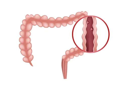
• Lower GI Tract: Colorectal polyps, colorectal adenocarcinoma, diverticular disease, inflammatory bowel disease (IBD), or vascular malformations (angiodysplasias).
• Small Bowel: Angiodysplasias, Crohn’s disease, subclinical malabsorption syndromes, or small bowel tumours.
Clinical Algorithm: Deciding the Next Diagnostic Step
When evaluating a patient with confirmed iron deficiency anaemia where non-GI causes have been ruled out, a structured approach ensures timely diagnosis and treatment.
1. Initial Assessment & Celiac Screening
Before invasive procedures, review NSAID or anticoagulant use and screen for non-invasive causes like Celiac Disease using serology (Anti-tissue transglutaminase IgA), as villous atrophy directly impairs iron absorption.
2. Bidirectional Endoscopy (EGD + Colonoscopy)
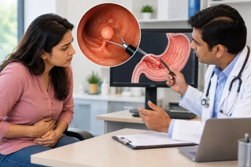
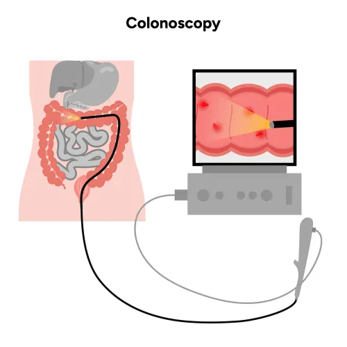
For most individuals with unexplained IDA, simultaneous or sequential upper endoscopy (EGD) and colonoscopy represent the gold-standard first-line diagnostic approach.
• EGD inspects the oesophagus, stomach, and duodenum, allowing for mucosal biopsies to evaluate celiac disease or H. pylori.
• Colonoscopy evaluates the large intestine and distal ileum to detect pre-cancerous polyps, colorectal cancer, or vascular malformations before symptoms like overt bleeding occur.
3. Video Capsule Endoscopy (VCE) for Obscure Bleeding
If bidirectional endoscopy yields no significant pathology but iron deficiency persists or requires recurrent transfusions, the source may lie in the middle GI tract (the small intestine). Video Capsule Endoscopy allows painless, non-invasive visualization of the entire small bowel to identify hidden angiodysplasias, small bowel ulcers, or rare tumours.
Why Early Screening Matters
1. Early Detection of Malignancy: Colorectal cancer and gastric malignancies often present with microcytic anaemia long before changes in bowel habits, abdominal pain, or overt haematochezia occur.
2. Preventing Complications: Identifying benign causes—such as large colon polyps or active peptic ulcers—allows for early endoscopic therapy, preventing severe acute bleeding or progression to advanced disease.
3. Restoring Quality of Life: Addressing the root cause ensures long-term resolution of fatigue, reduced exercise tolerance, and cognitive brain fog associated with chronic anaemia.
A Note for Referring Physicians & Patients
Treating iron deficiency with oral or intravenous iron addresses the symptom, but comprehensive gastroenterological evaluation addresses the cause. If you or one of your patients presents with unexplained low iron or anaemia without a clear source, early endoscopic evaluation can provide both a definitive diagnosis and peace of mind.
For consultations, endoscopic evaluations, or clinical inquiries, visit www.drpravishghurde.com.
Keywords